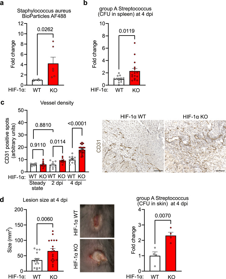
Fig. 4. HIF-1α in NK cells is required to control bacterial skin infections.
a Flow cytometry analysis of the spleen from WT and HIF-1α KO animals for Staphylococcus aureus bioparticles labelled with AlexaFluor 488 injected to the injury site, pooled of 3 experiments, two-tailed Student’s t test. b Colony forming units (CFU) of group A Streptococcus (GAS) in the spleen from WT and HIF-1α KO animals at 4 days post infection (dpi), pooled data of 4 experiments, (n = 13 for WT and n = 16 for HIF-1α KO), two-tailed Student’s t test. c Left, quantitative analysis of CD31-positive endothelial cells in skin lesions at 2- and 4- dpi with GAS from WT (steady state, n = 9; day 2, n = 8; day 4, n = 12) and HIF-1α KO animals (steady state, n = 9; day 2, n = 9; day 4, n = 20), pooled data of 3 experiments, one-way ANOVA test. Right, representative images of CD31 immunohistochemistry on skin wounds. Scale bar, 100 μm. d Left, quantification of skin lesion size at 4 dpi with GAS in WT and HIF-1α KO animals. Pooled data of 4 different experiments, (n = 16 lesions for WT and n = 19 for HIF-1α KO), two-tailed Student’s t test. Middle, macroscopic images of GAS skin lesions in WT and HIF-1α KO animals at 4 dpi. Right, GAS CFU in skin lesions from WT and HIF-1α KO animals at 4 dpi, (n = 4), two-tailed Student’s t test. Data are mean values ± SEM.

