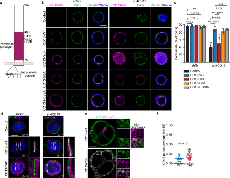
Fig. 7. The intracellular domain of CD13 is required to maintain apical-basal polarity.
a Schematic diagram showing mutation sites at intracellular and peptidase domain of CD13. b Confocal images of CD13-V5 (magenta) and Par6 (green) showing the rescue phenotype in wildtype and different CD13 mutants of shScr and shCD13 Caco-2 cysts. c Quantification of the percentage of Caco-2 spheroids with internally localized Par6 in wildtype (shScr, n = 515; shCD13, n = 292), CD13-Y6F (shScr, n = 462; shCD13, n = 433), CD13-S8A (shScr, n = 541; shCD13, n = 333), and CD13-H388A (shScr, n = 452; shCD13, n = 336) of shScr (n = 428) and shCD13 (n = 492) Caco-2 cysts. Dots represent mean values ± SD from each of 10 experiments. p-values were calculated by ANOVA with Tukey’s posthoc test for multiple comparisons. d Images for CD13-V5 (magenta) and Par6 (green) showing the rescue phenotype in wildtype and CD13-Y6F mutant of shScr and shCD13 Caco-2 in 2-cell structures. Numbers in the lower right indicate the fraction of cells analyzed with Par6 localized between the nuclei. e Caco-2 cells expressing CD13-WT or CD13-Y6F were immunostained for CD13-V5 (magenta) and Calreticulin to label the endoplasmic reticulum (ER) (green). f Quantification of the proportion of CD13 puncta that overlap with sites of ER-positivity. Dots represent individual Caco-2 structures from a single experiment. p-values were calculated with an unpaired two-tailed student’s t-test. Images are representative from two (b, d) independent experiments. Bars: b, 50 μm; d, e, 10 μm.

