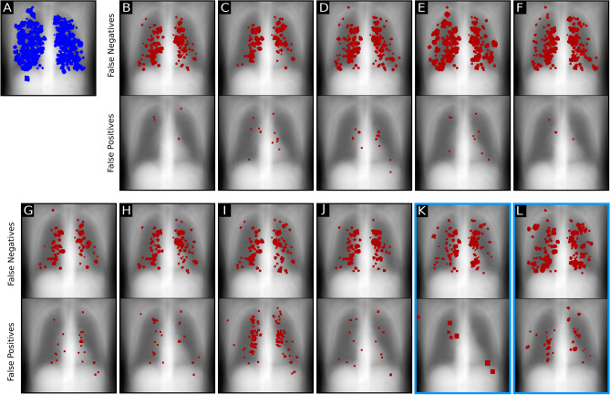
Figure 2.
Localization of false negative and false positive predictions in the reader study. Backgrounds were determined by averaging over all reader study radiographs. (A) All inserted nodules of different sizes in all radiographs marked blue. (B–J) False negative and false positive predictions by reader. (K) Location of false negative predictions of RetinaNet and false positive predictions of RetinaNet. (L) False negative predictions of U-Net and false positive predictions of U-Net.

