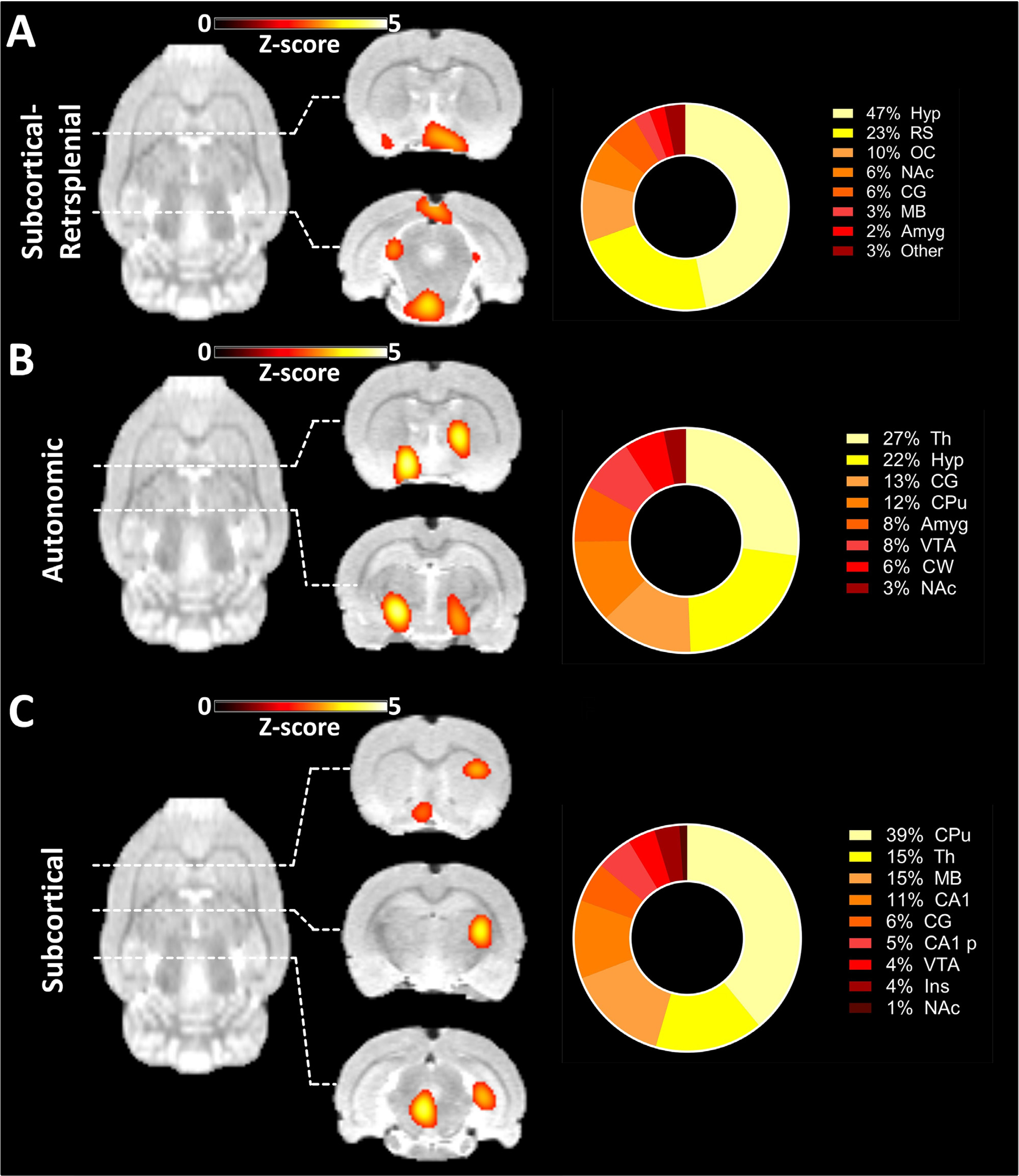
Fig. 7.

Components found exclusively in [18F]FDG-PET-derived ICA. (A) Component comprising the Hyp, RS, OC and other subcortical and posterior areas along with the regional quantification of the derived signal. (B) Component mainly composed of Th, Hyp and other subcortical areas along with the regional quantification of the derived signal. (C) Component comprising multiple subcortical areas along with the regional quantification of the derived signal. The results are reported at group-mean level (n = 30). ICA = independent component analysis. For a list of abbreviations of all regions, please refer to Supplementary Table 1.
