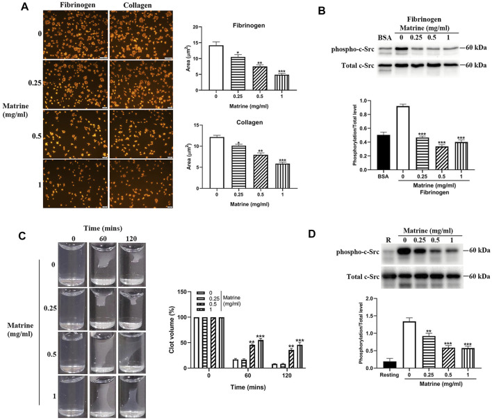
FIGURE 3.
Platelet spreading and clot retraction. After matrine treatment, platelets were placed on fibrinogen or collagen coated glass coverslips and allowed to spread at 37°C for 90 min followed by staining with Alexa Fluor-546-labelled phalloidin (A) (mean ± SE, n = 3) or measurement of c-Src phosphorylation by western blot (mean ± SD, n = 3) (B). Clot retraction was initiated in matrine-treated platelets in the presence of 2 mM Ca2+ and 0.5 mg/ml fibrinogen after addition of thrombin (1 U/ml). Images were captured every 30 min (mean ± SD, n = 3) (C). Under clot retraction condition, the phosphorylation level of c-Src was measured by western blot and represented as a ratio relative to the total level (mean ± SD, n = 3) (D). BSA: bovine serum albumin. Compared with 0, * p < 0.05; **p < 0.01; ***p < 0.001.

