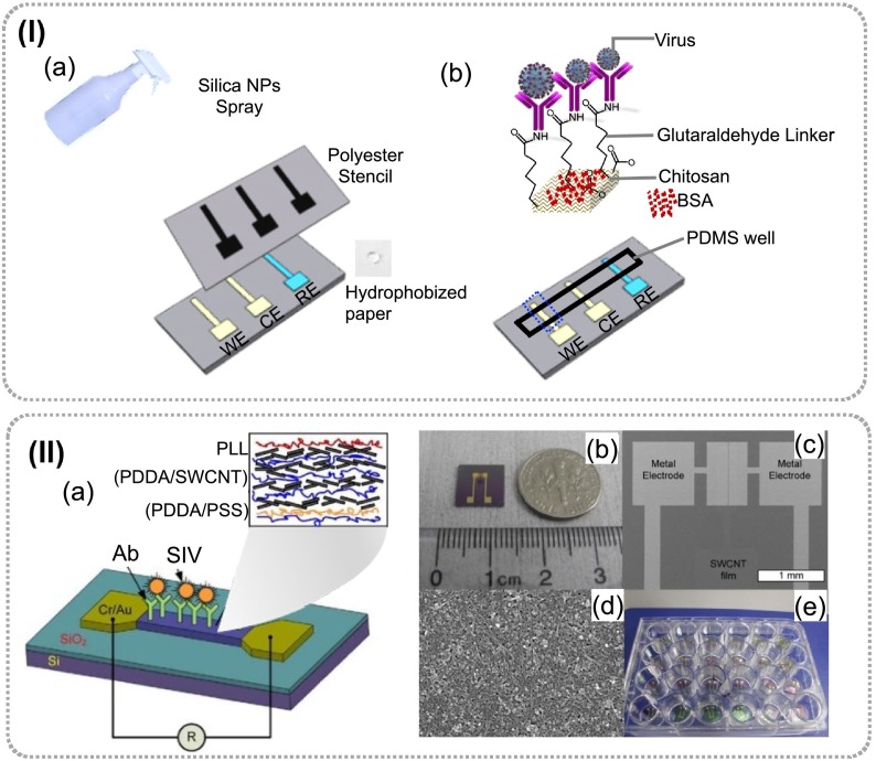
Fig. 23.
Schematic diagram of; (a) fabricating a hydrophobic paper with the help of a glass vaporizer and a polyester stencil, and (b) Functionalization of the CNT–chitosan modified working electrode for H1N1 virus detection and three electrode paper-based electrochemical immunosensor embedded with a PDMS well carrying electrolyte [197]. (II) Schematic shows the developed CNTs based immunochips: (a) immunoassay showing thin film formed by self-assembled CNTs random network and capturing of virus on modified electrode surface, (b) an optical immunoassay CNT chip, (c) SEM-images showed metal electrode (Cr/Au) pattern, (d) A thin file of multilayered SWCNTs between the two electrodes and random network of SWNCTs in SEM, and (e) a microtiter plate in which immunochips were sorted for use in immunoassay [90].

