
Figure 1.
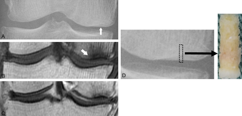
Images and histology of a 68-year-old female (case 2). A. Anteroposterior radiograph of the right knee shows flattening and slight sclerotic change (arrow) of the medial condyle. B. T1-weighted MRI coronal slice shows diffuse low intensity at the medial femoral condyle, with an associated lower intensity line at 5.0 mm from the articular surface (arrow). C. Gadolinium-enhanced T1-weighted MRI shows enhancement of the low intensity areas. D. An osteochondral pillar obtained from the affected area at the medial condyle. Biopsy diameter is 2.4 mm and depth is 13 mm.