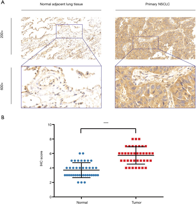
Figure 5.
Expression of ERβ and evaluated via immunohistochemical analyses of primary NSCLC tissue and normal adjacent lung tissue. (A) Representative IHC staining images from paired human primary NSCLC tissue and their normal adjacent lung tissue for ERβ. (B) Quantification data of IHC score for 39 paired primary NSCLC tissues and normal adjacent lung tissues. ****P<0.0001, t-test. ERβ, estrogen receptor beta; NSCLC, non-small cell lung cancer; IHC, immunohistochemistry.

