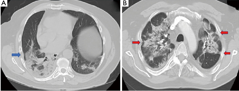
Figure 1.
Axial chest imaging with pleural changes often seen in early disease. (A) CT chest in a 70-year old male obtained 5 days following hospitalization showed pleural thickening (blue arrow) adjacent to subpleural parenchymal infiltrate. (B) CT scan of the chest in a 55-year old male seven days after admission demonstrated bilateral pleural retraction (red arrow).

