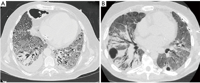
Figure 2.
Pleural involvement in progressive late disease. (A) Axial CT scan of the chest four weeks following hospitalization in a 60-year old man without any comorbidities revealed a small right sided pleural effusion. Advanced fibrotic changes with traction bronchiectasis was present bilaterally. Parenchymal destruction with pneumatocele formation was noted in the anterior right chest. (B) CT scan of the chest in a 51-year old male 30 days after hospitalization demonstrated bilateral pneumatocele formation. The patient developed a left sided pneumothorax and underwent small bore chest tube placement. Advanced pulmonary parenchymal changes were also seen.

