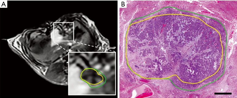
Figure 4.
Magnetic resonance imaging (MRI) and histology-based irreversible electroporation (IRE) and reversible electroporation (RE) areas. (A) Representative postcontrast T1-weighted MRI where the reversibly electroporated zone is outlined in yellow and the peripheral reversibly electroporated zone in green. (B) H&E-stained histology slide, corresponding regions were demonstrated in which the necrotic (irreversible) center is marked in yellow and the enhanced rim is emphasized in green. [Adapted from Figini et al. (87). Copyright 2018 by John Wiley & Sons Ltd.].

