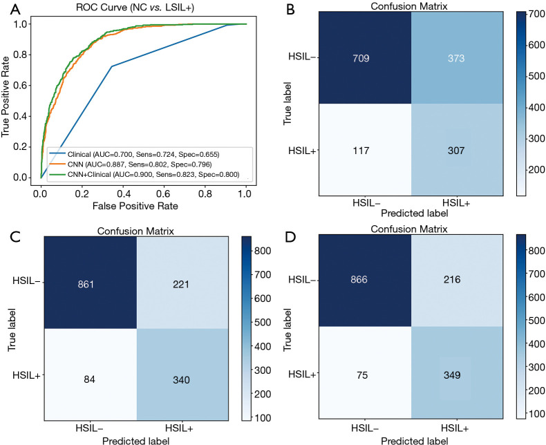
Figure 4.
Classification results of HSIL− vs. HSIL+. (A) ROC curves; (B) confusion matrix of the model based on clinical features; (C) confusion matrix of the model based on ResNet50; (D) confusion matrix of the model based on the combination of ResNet50 and clinical features. HSIL, high-grade squamous intraepithelial lesions; ROC, receiver operating characteristic; ResNet, residual neural network.

