Figure 4.
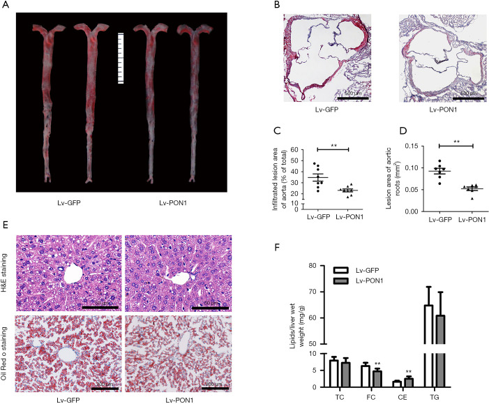
PON1 overexpression attenuates atherosclerosis and hepatic steatosis in Scarb1−/− mice. (A) Representative images of atherosclerotic lesions by en face aortas Oil Red O-staining. (B) Atherosclerotic lesions were examined by Oil Red O-stained cross-sections of the aortic root (8µm serial sections). Scale bars: 500 µm. (C) Relative quantification of lesion area in the Lv-GFP and Lv-PON1 mice, data are presented as the percentages of en face aortic area. n=8 per group. (D) The cross-sectional lesion areas of the aortic roots were quantified. The data are presented as the lesion areas (mm2). n=7 per group. (E) Hepatic lipid levels were analyzed by hematoxylin and eosin (H&E) or Oil Red O staining. Scale bars: 50 and 100 µm, respectively. (F) The contents of TC, FC and TG were determined using isopropanol as described in Methods. n=8–9 per group. The measurement data were presented as mean ± standard error of the mean. Data between the two groups were compared by unpaired Student’s t-test. **P<0.01, versus Lv-GFP group.

