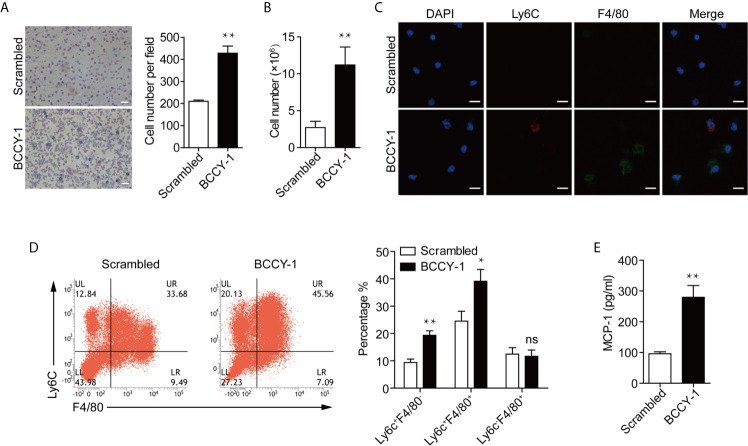
Figure 3.
Effects of BCCY-1 on monocyte recruitment in vivo. Monocyte recruitment into the peritoneum induced by peptide BCCY-1 (10 mg/kg) post i.p. injection was analyzed. The scrambled version of BCCY-1 served as a negative control. (A) Wright-Giemsa stain of peritoneal lavage fluid after BCCY-1 or Scrambled peptide administration at 24 h. Bar=200 μm. The nucleated cell number per field is shown as a bar graph in the right panel. **p < 0.01. (B) Cell number of peritoneal lavage cells after BCCY-1 or Scrambled peptide administration at 48 h. **p < 0.01. (C) Representative confocal images of peritoneal lavage cells after the indicated treatment are shown. Nuclei were stained with DAPI (Blue: DAPI; Red: Ly6C; Green: F4/80). Bar=20 μm. (D) Monocyte recruitment at 48 h post BCCY-1 injection was analyzed by flow cytometry using the following markers: Ly6C (monocytes), F4/80 (macrophages). Quantification of monocyte/macrophage percentage is shown as a bar graph in the right panel. ns p > 0.05, *p < 0.05, **p < 0.01. (E) Peritoneal lavage fluids at 48 h after the indicated treatment were analyzed for MCP-1 production by ELISA. **p < 0.01.

