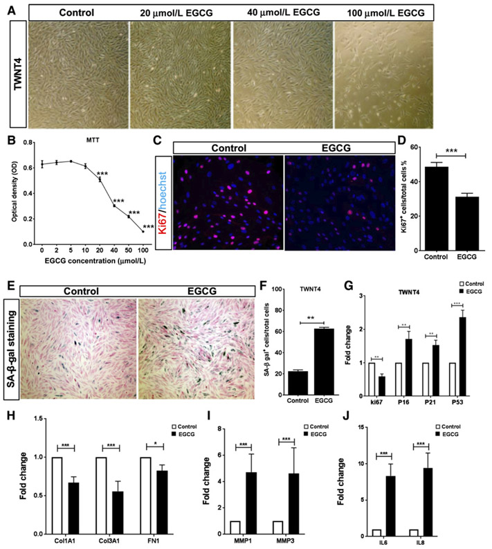
Figure 4.
EGCG induces SASP in human HSCs cell line in vitro. TWNT4 cells were treated with different concentration of EGCG (0, 2, 5, 10, 20, 40, 50, and 100 μmol/L) for 72 hours. A, Representative bright field pictures of TWNT4 cells treated with 20, 50, and 100 μmol/L of EGCG. B, MTT cell proliferation assay in TWNT4 cells, 72 hours after treatment with EGCG. C and D, Immunofluorescence staining of Ki67 (red) and DAPI (blue) on TWNT4 cells after 72 hours treatment with 20 μmol/L EGCG. Untreated cells were considered as control, 2,000 cells were counted in each group; ***, P < 0.001 compared with control. E and F, SA-β-gal staining (turquoise blue) of TWNT4 cells after 72 hours treatment with 20 μmol/L EGCG. Untreated cells were considered as control, 2,000 cells were counted in each group; **, P < 0.01 compared with control. G-J, RNA expression of senescence and SASP markers in TWNT4 cells after 72 hours treatment with 20 μmol/L EGCG. Untreated cells were considered as control. N = 4 replicates (*, P < 0.05; **, P < 0.01; ***, P < 0.001).

