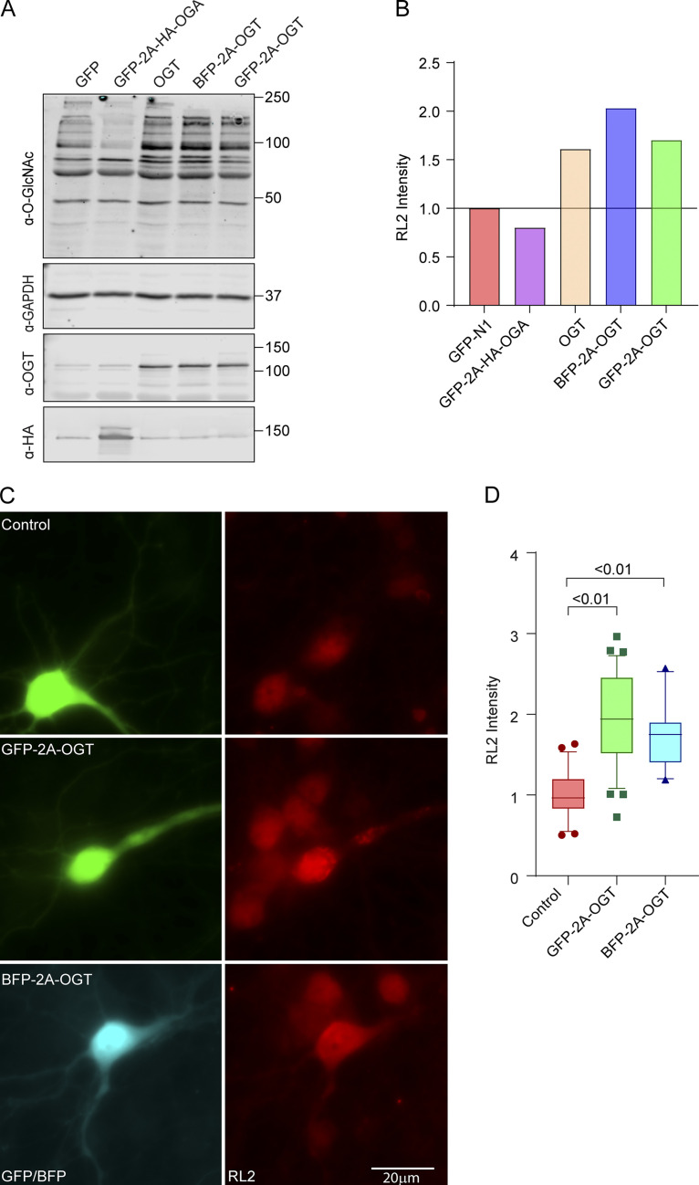
Figure S1.
Verification of OGT and OGA constructs in COS-7 cells (related to Fig. 1). (A and B) Construct verification in COS-7 cells. COS-7 cells were transfected with OGT or OGA constructs and lysed after 2 d. (A) Representative Western blot of lysates probed for OGT, OGA (HA), GAPDH, and O-GlcNAcylation (with antibody RL2). Molecular weights (in kD) are indicated on the right. (B) O-GlcNAcylation was quantified by normalizing the intensity of RL2 staining (full lane) to that of the GAPDH band. Values were then expressed relative to those cells expressing only GFP. Each bicistronic construct was almost fully cleaved to release OGA or OGT. Expression of OGT increased, and OGA decreased, O-GlcNAcylation levels relative to the GFP (control)-expressing cells. (C and D) Construct verification in neurons. GFP-2A-OGT, BFP-2A-OGT, or GFP constructs were expressed in rat hippocampal neurons for 3 d. (C) Representative images of neurons stained with the O-GlcNAc antibody RL2. (D) To quantify the RL2 intensity, the BFP or GFP signal was used to mask the cell bodies, and total RL2 intensity inside the mask was measured and normalized to the control. n = 25–30 cells per condition from 3 independent animals.

