Figure S5.
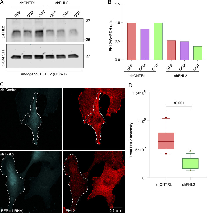
Validation in COS-7 cells of shRNA and antibody against FHL2 (related to Fig. 5). (A and B) COS-7 cells were transfected with OGT, OGA, and GFP along with an shRNA to FHL2. The cells were then lysed, and the lysates were probed for FHL2 and GAPDH to confirm FHL2 knockdown (A). In all cases, the shRNA against FHL2 was effective in reducing the levels of FHL2 (as compared with GAPDH, loading control) by >50% (B). (C and D) Validation of antibody against FHL2 used for immunocytochemistry in COS-7 cells. The FHL2 shRNA plasmid was modified to coexpress BFP to mark the transfected cells. Cultures expressing the FHL2 shRNA or a control shRNA were fixed and stained with the antibody against FHL2 (C). Left panels indicate BFP-tagged cells; right panels show the FHL2 staining as a sum projection of confocal slices. (D) The total intensity of FHL2 staining was quantified in the transfected cells. n = 10–15 cells from 3 independent transfections. Quantifications are represented as box-and whisker plots. The line indicates the median, the box indicates the interquartile range, and whiskers indicate the 10th and 90th percentiles. Outliers are represented as individual dots and are included in all statistical calculations. P value is from two-tailed unpaired t test with Welch’s correction.

