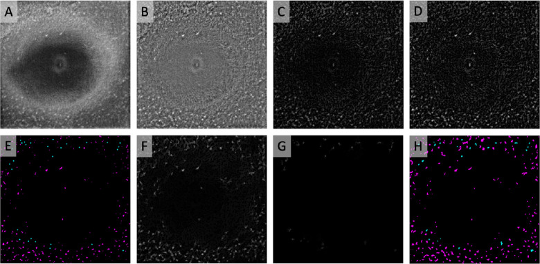
Figure 2.
Semiautomated binarization protocol to identify MLCs. (A) Registered and averaged 3-µm OCT slab containing MLCs. (B) Background-flattened MLC layer OCT slab. (C) Background-subtracted slab from B. (D) MLC signal from C was compensated by multiplying against an averaged, inverted slab composed of the blurred MLC layer OCT and RNFL layer (not shown). (E) Automated (magenta) seeds were generated from the compensated image using MaxEntropy binarization. Manual seeds (cyan; enlarged for clarity) were placed on cells that were not identified by automated binarization. (F) Cells filled in by morphological dilation from automated seeds in E. (G) Manually seeded lower-intensity cells missed by automated seeding were filled in with a separate morphological dilation step. (H) Combined image containing binarized cells from automated seeding (magenta) and manual seeding (cyan).

