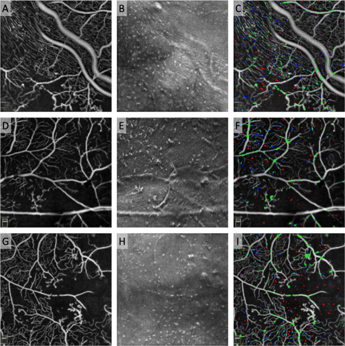
Figure 7.
MLCs clustered around but not in large areas of ischemia outside the macula. Top row: Eye with severe NPDR imaged in the inferior quadrant near the vascular arcades. Middle and bottom rows: Eyes with PDR also imaged outside the macula. The first column shows the aligned and averaged OCTA of the SCP. The second column shows the aligned and averaged 3-µm OCT slab located from 0 to 3 µm above the ILM with hyperreflective dots representing MLCs. The third column shows the distribution of pseudo-colored MLCs overlaid on the OCTA image. Green MLCs colocalized with SCP vessels or to both blood vessels and the perivascular space, blue MLCs are located in the perivascular area within 3 pixels (∼30 µm) of the nearest vessel, and red MLCs are located in the ischemic area more than ∼30 µm from the nearest vessel or in both the ischemic area and perivascular space.

