
Fig. 2.
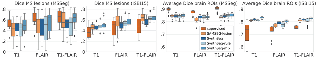
(a) Box plots of the cross-validation Dice scores for the MS lesions and (c) for the average over 12 brain ROIs: cerebral cortex and white matter, lateral ventricle, cerebellar cortex and white matter, thalamus, caudate, putamen, pallidum, brainstem, hippocampus, and amygdala. (b) and (d) show the results obtained when training on MSSeg and testing on ISBI15.