Figure 3.

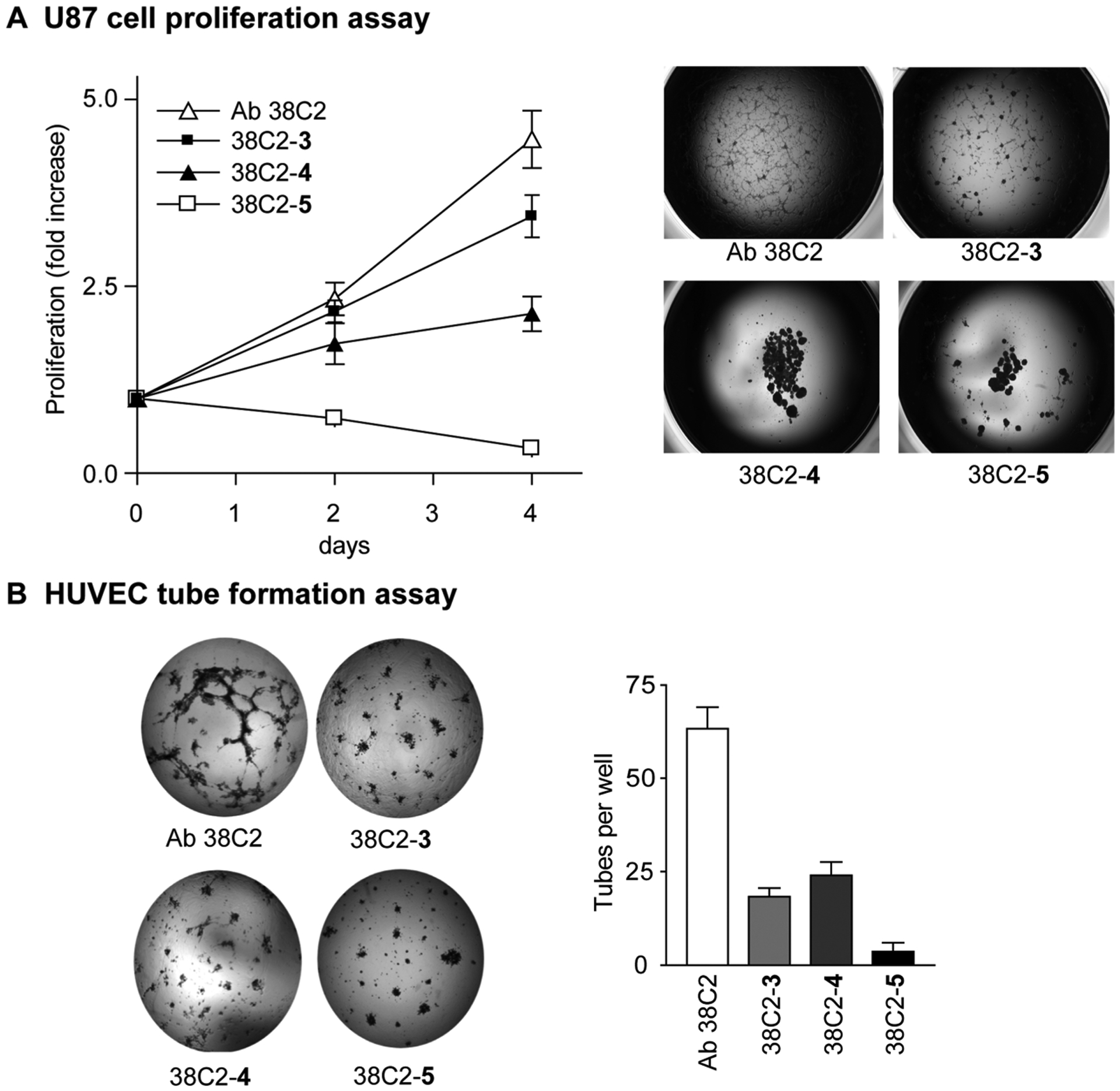
In vitro cell proliferation and tube formation assays. (A) A line graph showing the proliferation of human U87 cells treated with Ab 38C2, cpAbs 38C2-3 and 38C2-4, and cp-bsAb 38C2-5 (left) (38C2-5 vs 38C2-3, p < 0.001; 38C2-5 vs 38C2-4, p < 0.001). Microscope image showing morphology of the human U87 cells on day 6 after cells were treated with Ab 38C2 or the chem-Abs (right). (B) 38C2-3, 38C2-4, and 38C2-5 inhibit HUVEC tube formation in vitro (left). Tube formation was quantified by comparing pixel tube number in each image by using the NIH Image program (right). The experiments were repeated three times. (38C2-5 vs 38C2-3, p < 0.001; 38C2-5 vs 38C2-4, p < 0.001.) Data are represented as mean ± SEM.
