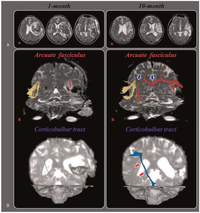
Figure 1.
(A) Brain MR images at 1 month after onset show an infarct in the left middle cerebral artery territory, hemorrhagic transformation and subfalcine herniation. Brain MR images at 10 months after onset reveal leukomalactic lesions in the left fronto-parieto-temporo-occipital areas. (B) Results of diffusion tensor tractography (DTT) for the arcuate fasciculus (AF) and corticobulbar tract (CBT). On one-month DTT, the discontinuation of the left AF and severe narrowing of the right CBT are observed. By contrast, on 10-month DTT, the left AF is connected to opposite AF by a new tract that passed through the splenium of corpus callosum (blue arrows) and the right CBT become thicker (red arrows).

