. 2020 Nov 3;2(1):192–205. doi: 10.1039/d0cb00157k

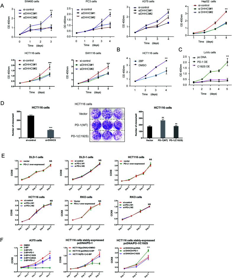
Fig. 6. Targeting palmitoylation suppressed tumor-intrinsic PD-1 functions. (A) CCK8 proliferation assay showing that the proliferation of cancer cells treated with siRNAs for DHHC9 is decreased. *** P < 0.001, ** P < 0.01, ANOVA test. n = 3 independent experiments. (B) Treatment of HCT116 cells with 2-BP decreases the proliferation of cells as detected by CCK8 assay. (C) Overexpressing the C192S mutant (C192S OE) decreases cell proliferation compared to WT PD-1 (PD-1 OE) in LoVo cells. (D) Overexpressing WT PD-1 (PD-1 OE) increases anchorage-free colony formation assay (statistics shown on the right, PD-1 C192S compared to WT, and WT compared to Vector. ** P < 0.01, ANOVA test. n = 3 independent experiments) and targeting DHHC9 decreases it compared to control conditions (statistics shown on the left, *** P < 0.001, ANOVA test. n = 3 independent experiments). (E) CCK8 proliferation assay showing that the proliferation of cancer cells was not affected by the transfection of siRNAs for PD-L1 or PD-1 plasmid. NS, P > 0.05, ANOVA test. (F) CCK8 proliferation assay showing that PD-1 and its C284A mutant but not its C192S mutant or PD-L1 could rescue the effect of 2-BP or si-DHHC9 on tumor cell proliferation. *** P < 0.001, NS, P > 0.05, ANOVA test. n = 3 independent experiments.

Fig. 6