
Fig. 3. On microscopy, microlithiasis is often defined as a tiny stone with a diameter < 3 mm, which cannot be crushed by digital compression.
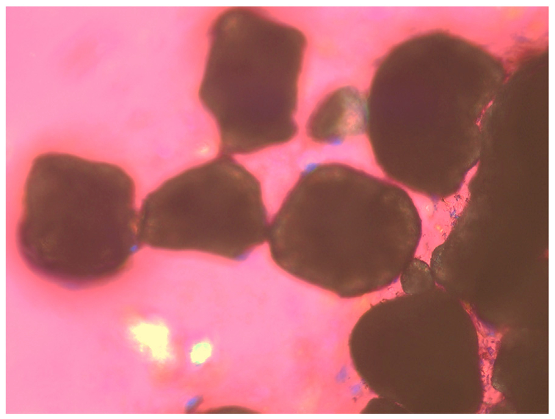
Under phase contrast and polarizing light microscopy, microlithiasis always display rounded contours and black centers from light scattering/absorption. Original magnification is × 100 by polarizing light microscopy.