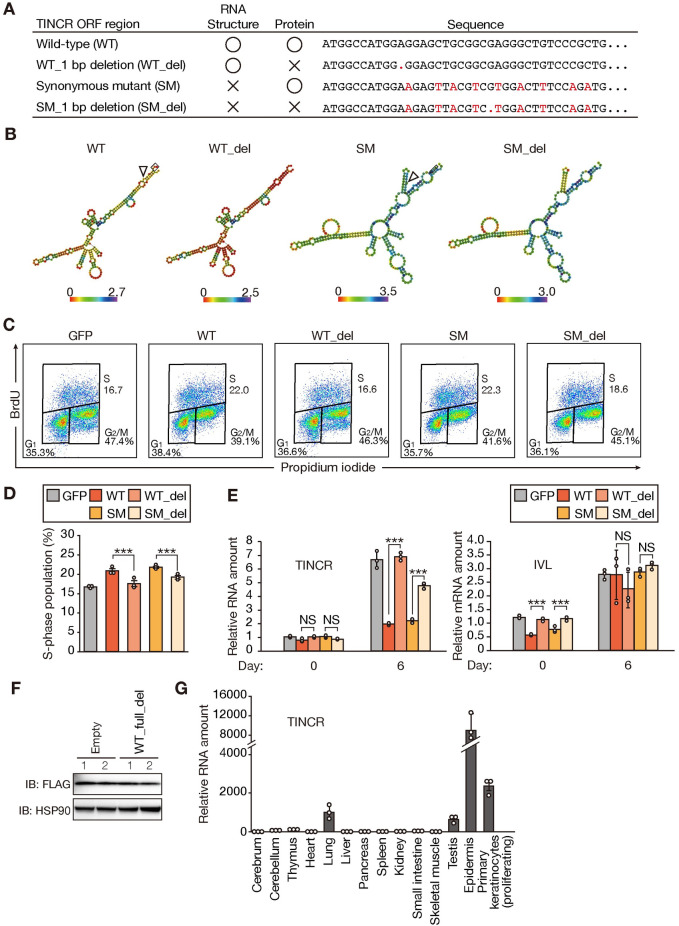
Fig 3. TUBL expression promotes proliferation of mouse primary keratinocytes in a manner independent of the secondary structure of TINCR RNA.
(A) Summary of mutations introduced into the mouse TUBL ORF. (B) Predicted secondary structure and minimal free energy for WT, WT_del, SM, and SM_del. (C, D) Flow cytometric traces (C) and quantification (D) of BrdU incorporation for mouse primary keratinocytes stably expressing either GFP or WT, WT_del, SM, or SM_del forms of TINCR. Data in (D) are means ± SD (n = 3 independent experiments). ***p < 0.005 (Student’s t test). (E) RT-qPCR analysis of TINCR and involucrin (IVL) mRNA abundance in mouse primary keratinocytes stably expressing either GFP or WT, WT_del, SM, or SM_del forms of TINCR and subjected to calcium-induced differentiation in vitro for 0 or 6 days. Data are means ± SD (n = 3 independent experiments). ***p < 0.005, NS (Student’s t test). (F) Immunoblot analysis of HEK293T cells transiently transfected both with an expression vector for the mouse TUBL ORF with a COOH-terminal FLAG epitope tag and with either an expression vector for an almost full-length form of mouse TINCR with a 1-bp deletion in the TUBL ORF (WT_full_del) or the corresponding empty vector. Two replicates (lanes 1 and 2) are shown. (G) RT-qPCR analysis of TINCR in adult mouse tissues and actively proliferating primary mouse keratinocytes. Data are means ± SD (n = 3 independent experiments).

