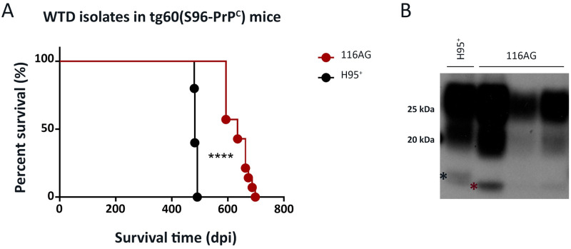
Fig 2. Transmission of the 116AG isolate to transgenic tg60 mice expressing S96-deer PrP.
Tg60 mice, expressing S96-deer PrP at 0.7fold, were inoculated with 20 ul of 1% (w/v) brain homogenate containing 116AG or H95+ prions. (A) A Kaplan-Meier curve depicting the survival times of tg60 mice inoculated with 116AG or H95+ strains. ****p <0.0001, statistical differences were evaluated using a log-rank (Mantel-Cox) test. (B) Western blot of brain homogenates from mice inoculated with, H95+ (lane 1) or 116AG (lanes 2–4) prions after PK digestion. PrPres was detected using the anti-prion monoclonal antibody BAR224. The asterisks show the migration profile, slow (black asterisk) and rapid (red asterisk), of the non-glycosylated bands.

