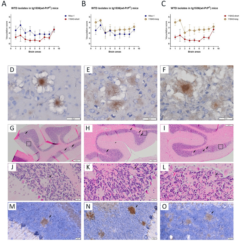
Fig 5. Strain characterization of tg1536+/+ mice inoculated with Wisc-1 and 116AG isolates.
Neuropathological profiles of brains of tg1536+/+ animals inoculated with 10−4 dilution of Wisc-1 and 116AG prions, respectively, were determined. Brain vacuolation profiles comparing (A) 116AG-short and Wisc-1 inoculated mice, (B) 116AG-long and Wisc-1 inoculated mice, and (C) 116AG-short and -long inoculated mice. The y-axis represents vacuolation scores on a range of 0 to 5 in terms of spongiform changes (0 is none and 5 is severe). The x-axis represents the brain areas scored (1 frontal cortex, 2 basal ganglia, 3 parietal cortex, 4 hippocampus, 5 thalamus, 6 hypothalamus, 7 midbrain, 8 cerebellum, 9 medulla/pons). The scoring was performed in a blinded manner at least six times (range 6–8). Each curve represents each group, and each data point is the mean vacuolation score ± standard deviation. (D-F) Presence of florid plaques after PrPSc immunostaining using 12B2 mAb in the frontal cortex brain sections of mice inoculated with (D) Wisc-1, (E) 116AG-short and (F) 116AG-long prions. (G-O) Presence of unicentric plaques in the granular layer of the cerebellum of mice inoculated exclusively with 116AG-long. (G-I) H&E staining of three representative brains from mice inoculated with serially diluted brain homogenate of 116AG-long prions (10−3, 10−4 and 10−5). The unicentric plaques are indicated with black arrows and black squares. (J-L) H&E staining showing unicentric plaques highlighted by squares at a higher magnification. (M-O) PrPSc immunostaining using 12B2 mAb of the unicentric plaques in the granular layer of the cerebellum at a higher magnification. Scale bars indicate 20 μm and 50 μm for high magnification and 200 μm.

