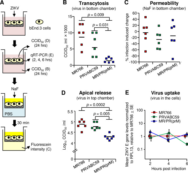
Fig 6. Viral transcytosis and uptake in bEnd.3 cells.
(A) In vitro Transwell setup. Mouse endothelial cells (bEnd.3) were seeded onto the luminal side of the Transwell insert. After the bEnd.3 monolayers reached confluence, MR766, PRVABC59 or MR/PR(prM) viruses were added to the top well at a MOI of 1. After 2, 4 and 6 hrs, viral RNA levels in the cells were determined by qRT-PCR, and after 24 hrs, virus titers in the upper and lower chambers were determined by CCID50 assays. After collecting the culture medium from the upper and lower chambers at 24 hrs, NaF was added to the top wells and PBS to the bottom chambers; after 30 min, samples from the bottom chambers were analyzed by fluorometer. (B) ZIKV levels in the lower chambers. The medium in the lower chamber was collected and virus titers were determined. Kolmogorov-Smirnov tests were used for statistics. Data were obtained from two independent experiments. The horizontal line indicates the mean. (C) The percent of virus-induced permeability. The percent virus-induced permeability was calculated relative to a linear standard curve where “no virus control” was 0% and “no cell control” was 100%. T-test was used for statistical analysis. (D) ZIKV levels in the upper chambers. The medium in the upper chambers was collected and virus titers were determined. Kolmogorov-Smirnov tests were used for statistical analyses. (E) Viral uptake by bEnd.3 cells. bEnd.3 cells were inoculated at a MOI of 1 and incubated for 2, 4 and 6 hrs. The cells were washed three times with PBS, trypsinized, washed three times with PBS, and dissolved in TRIzol (n = 4–9 replicates per virus per time point). Uninfected bEnd.3 cells were used as controls (n = 3). ZIKV RNA levels in the cells were determined by qRT-PCR and normalized to RPL13 mRNA levels and graphed relative to MR766 for each time point. Data were obtained from three independent experiments.

