. 2020 May 27;1(2):42–55. doi: 10.1039/d0cb00041h

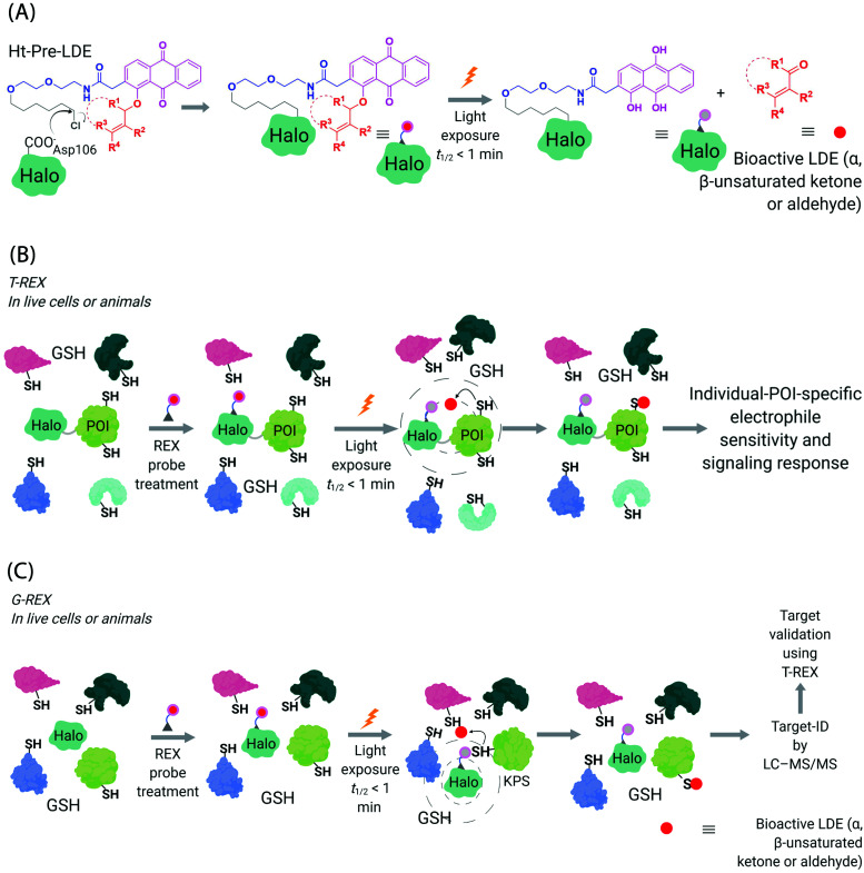
Fig. 3. Functional subproteomics mapping and on-target signaling-interrogation techniques, G-REX and T-REX. (A) G-REX maps functional subproteomes primed to sense lipid-derived electrophiles (LDEs) and related covalent drugs, whereas T-REX functionally validates and evaluates individual-POI-specific covalent-ligand sensitivity and signaling responses.82 The figure shows chemistry underlying photocaged electrophiles (Ht-Pre-LDEs) common to both G-REX and T-REX methods. Halo in G-REX (or Halo-POI in T-REX) expressed in cells or animals covalently binds Ht-Pre-LDE, following cell/animal incubation with designated Ht-Pre-LDE in culture media (typically at 1 μM overnight, or at 10 μM over 2–3 h). Following washout of excess, unbound Ht-Pre-LDE (step not shown in the figure), exposure of live cells/animals to light (365 nm, 5 mW cm−2 over 1–5 min) results in rapid liberation of LDE (t1/2 of photouncaging <1 min) within proximity of Halo in G-REX (or Halo-POI in T-REX). Concentration of LDE released is maximally stoichiometric to intracellular concentration of Halo/Halo-POI (which has been quantified to be <5 μM).70 (B) In T-REX, the protein of interest (POI) is fused to Halo. Cells/animals expressing Halo-POI are treated with designated Ht-Pre-LDE. Following rinsing cycles to remove the excess/unbound Ht-Pre-LDE, the system is exposed to low-energy UV light (see (A) legend). Providing the POI is a kinetically-privileged sensor (KPS) of the LDE in the vicinity of the POI in limited amounts, the LDE can be captured by the POI before it irreversibly diffuses away beyond the solvent shell of Halo-POI. See also Fig. 4B. Percentage LDE-occupancy of POI and the identity of LDE-sensing residue are assessed using previously published protocols. In parallel, T-REX set-up allows functional consequences of POI-specific covalent-ligand modifications to be assayed directly and precisely in intact cells/animals.65,66,70,71 (C) In G-REX, cells/animals expressing Halo (not fused to any protein) are treated with designated Ht-Pre-LDE and the subsequent steps remain the same as in T-REX (see Fig. 3B legend). The LDE (in alkyne-functionalized version) rapidly released in limited dosage in G-REX covalently tags the most kinetically-privileged native LDE sensors within the microenvironment of Halo in cells/animals. Following cell/animal lysis, endogenous KPSs covalently bound to the released ligand are enriched following Click-coupling to biotin-azide and streptavidin pulldown, and protein-ID is achieved using standard quantitative proteomics methods.70.

Fig. 3