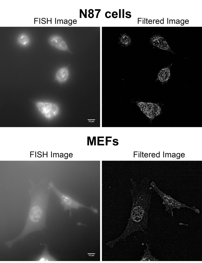
Figure 2. Examples of unfiltered and filtered FISH images produced by FISH-quant.
smFISH on human NCI-N87 gastric carcinoma cells (top row) and immortalized MEFs (bottom row) were performed using Stellaris probes against human HER2 mRNA. Imaging was performed on a Zeiss AxioObserver Z1 DuoLink dual camera imaging system equipped with Illuminator HXP 120 V light source, PlanApo 100x 1.4 NA oil immersion objective and Hamamatsu Flash 4 sCMOS cameras. For both cell types, 41 steps of 0.2 μm z-stack images were taken using a motorized XYZ scanning stage 130 x 100 PIEZO, and ZEN2 software at 0.0645 μm/pixel. Images show a maximum projection of middle z-sections. Note that the brightness of the MEFs FISH image (lower left) was increased compared to that of N87, to allow better visualization of the cells. See Videos 2-5 for the full z-stack of each image. Scale bars = 10 µm.

