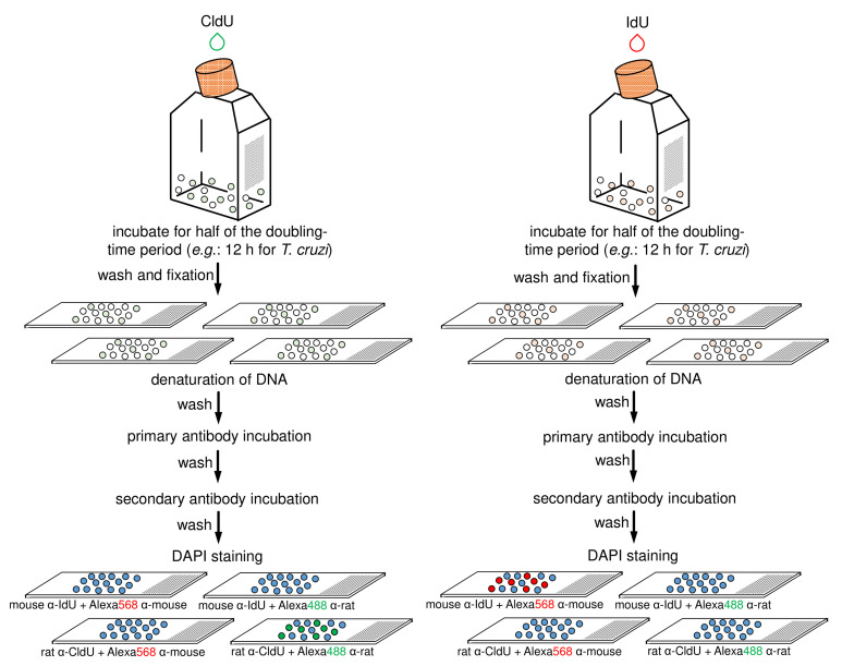
Figure 3. Schematic diagram representing the main steps of antibodies specificity control.
This control assay can be applied to any of the two previously strains (i.e., CL Brener and Y). The halogenated thymidine analogs (CldU and IdU) are added separately in each culture for 12 h. After that, each parasite-group (CldU-incorporated and IdU-incorporated) must be washed with 1x PBS, fixed, and aliquots distributed onto four slides (totalizing eight slides for the both groups). Next, each slide containing parasite cells must have their DNA denatured and should be then processed for the detection of the thymidine analogs using primary and secondary antibodies according to the specifications (on the scheme, see the specifications on the bottom of slides) (for details see previous Step B21). Finally, mounting medium with DAPI must be added in each slide and then sealed.

