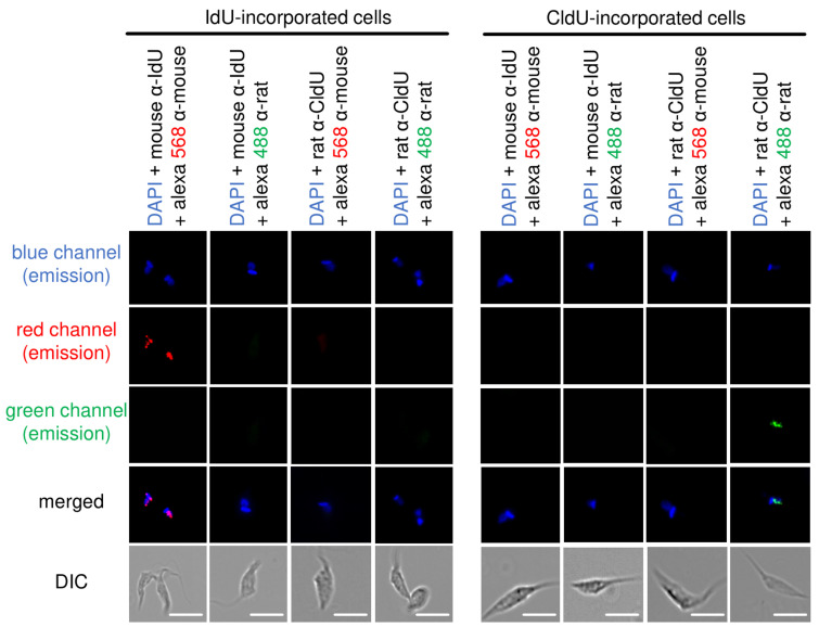
Figure 5. Antibodies specificity control.
Representative images of CL Brener strain show that the recognition of thymidine analogs is specific. CldU- and IdU-incorporated epimastigotes were added onto slides and processed for detection using mouse α-IdU + alexa fluor 568 (mouse), mouse α-IdU + alexa Fluor 488 (rat), rat α-CldU + alexa fluor 568 (mouse), and rat α-CldU + alexa fluor 488 (rat). We can observe complete absence of cross-reactions between primary and secondary antibodies in each fluorescence channel analyzed (blue, red and green). Images were captured randomly. This figure was adapted from Alves et al. (2018) . Scale bars = 10 μm.

