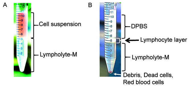
Figure 2. Density gradient separation procedure.

A. Before centrifugation, lymphoid cells are located in the cell suspension phase above the Lympholyte®-M. B. After centrifugation, lymphoid cells are concentrated in the interface between the DPBS buffer and the Lympholyte®-M. Red blood cells, dead cells and debris are found at the bottom of the tube.
