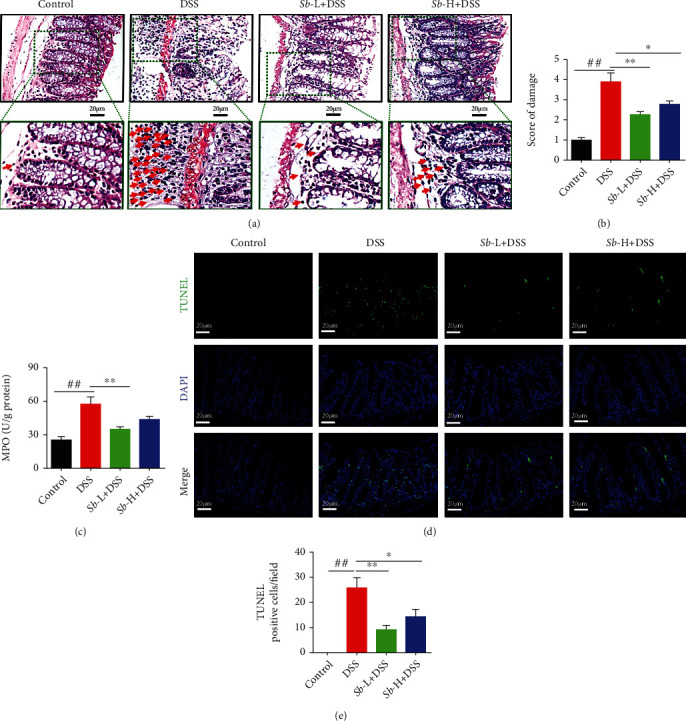
Figure 2.

S. boulardii alleviated pathological alterations in ulcerative colitis. (a) Representative images of colon injury shown by hematoxylin and eosin (HE) staining (Magnification: ×400; Scale bars: 20 μm). Arrows indicated the infiltration of neutrophils and monocytes in mucosa. (b) Histopathological score on colons. (c) Colonic MPO level. (d) Representative colon images of colon cell apoptosis. (e) Quantification of TUNEL positive cell number/field. N = 8. Values are presented as mean ± SEM. #P < 0.05 and ##P < 0.01 compared with the control group. ∗P < 0.05, and ∗∗P < 0.01 compared with the other groups. Sb: S. boulardii.
