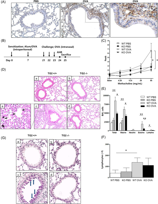
Figure 2.

TG2 expression and the effect of TG2 on lung inflammation and AHR following allergen challenge. TG2 expression in the lung of a murine asthma model (×400) (A). Experimental protocol of the study (B, n = 5–6 mice per group). Methacholine hyperresponsiveness was measured 24 h after the final intranasal OVA challenge. AHR was expressed as the enhanced pause (Penh) (C). H&E‐stained lung histology after allergen challenge from the mice of the different groups (a: WT PBS, b: KO PBS, c: WT OVA, d: KO OVA, e: black arrows indicate eosinophils, ×200) (D). The number of inflammatory cells, including eosinophils (E) and lymphocytes (F) in BAL fluid. PAS‐stained lung histology after allergen challenge from mice of the different groups (a: WT PBS, b: KO PBS, c: WT OVA, d: KO OVA, blue arrows indicate mucin secretion, ×400) (G). AHR, airway hyperresponsiveness; BAL, bronchoalveolar lavage; H&E, hematoxylin and eosin; KO, knockout; OVA, ovalbumin; PAS, periodic‐acid schiff; PBS, phosphate‐buffered saline; TG2, transglutaminase 2; WT, wild type. *p < .05, **p < .01.
