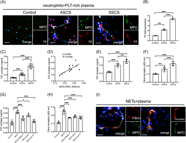
Figure 3.

NETs contribute to procoagulant activity by binding with coagulation factors through TF. (A) Control neutrophils were incubated with plasma from healthy subjects and patients with carotid stenosis. Neutrophils treated with ICH plasma expelled extracellular traps (arrowhead) stained with TF (red) and MPO (green). (B) Fold expression of TF mRNA in neutrophils incubated with plasma from controls and patients. (C) Detection of the TAT‐complex in plasma from healthy subjects and patients. (D) The TAT‐complex was positively correlated with H3cit‐DNA in plasma from symptomatic patients. Control plasma were incubated with NETs from controls and patients and the TAT‐complex (E) and fibrin formation (F) were detected. Control plasma was incubated with NETs (0.5 μgDNA/ml) then treated with DNase I, APC, and anti‐TF antibody in the inhibition assays. These inhibitors markedly decreased the thrombin (G) and fibrin formation (H). Colocalization of NETs and thrombin and fibrin were stained with MPO (green) and thrombin (red) and fibrin(red). The inset scale bar in A is 30 μm and I is 2030 μm. The results are expressed as the mean ± SD. APC, activated protein C; ICH, intracranial hemorrhage; NET, neutrophil extracellular traps; TF, tissue factor. *p < .05, **p < .01, ***p < .001 and ****p < .0001
