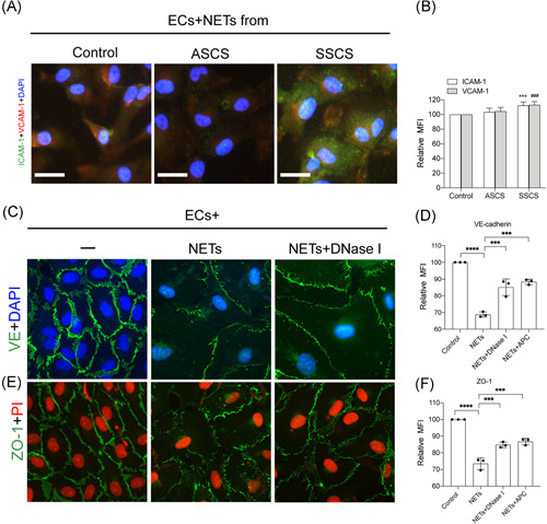
Figure 4.

NETs destroyed the endothelial barrier in symptomatic patients. ECs were incubated with control neutrophils and NETs from symptomatic and asymptomatic patients. (A) The expression of ICAM‐1 and VCAM‐1 from treated ECs in each group was analyzed through confocal microscopy. (B) The expression of ICAM‐1 and VCAM‐1 were quantified by MFI. The expression of VE‐cadherin (C) and ZO‐1 (E) in ECs treated with isolated NETs was analyzed through confocal microscopy. There was a decrease in the expression of VE‐cadherin and ZO‐1 when incubated with NETs (0.5 μgDNA/ml) and this cytotoxicity could be reversed by the NETs inhibitors, DNase I, and APC. The expression of VE‐cadherin (D) and ZO‐1 (F) were quantified by MFI. The inset scale bar in A, C, and E are 20 μm. The results are expressed as the mean ± SD. APC, activated protein C; EC, endothelial cell; ICAM‐1, intercellular adhesion molecule 1; MFI, mean fluorescence intensity; NET, neutrophil extracellular traps; VCAM‐1, vascular cell adhesion molecule‐1. *p < .05, **p < .01, ***p < .001 and ****p < .0001
