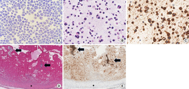
Fig. 2.
Cytological and histological images of peri-implant left breast seroma (A-C) and homolateral fibrous capsule (D, E). Papanicolaou stained cell smear (A, ×400) and hematoxylin and eosin-stained section of cell block (B, ×400) showed large pleomorphic cells with irregularly shaped nuclei and binucleated elements immunostained for CD30 (C, ×400). Histological examinations of the capsule revealed clusters of large atypical cells within a fibrinous exudate (black arrows) on the inner surface of the capsule (black asterisk) (D, H&E, ×100). The anaplastic cells were highlighted by CD30 stain (E, ×100).

