Fig. 6.

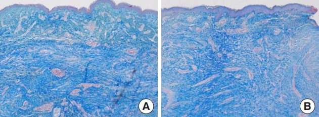
Histological findings. Masson’s trichrome staining (×40) revealed poorly structured collagen fibers on the untreated side (A) and increased dermal collagen on the treated side (B).

Histological findings. Masson’s trichrome staining (×40) revealed poorly structured collagen fibers on the untreated side (A) and increased dermal collagen on the treated side (B).