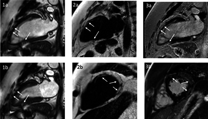
Fig. 4.
A 45-year-old patient with LGMD2I and normal LV function (LVEF 57%). SSFP cine in a two-chamber view in end-diastole (1a) and end-systole (1b) with no wall motion abnormality, but with detectable myocardial structure abnormality (arrows). A 2-chamber view and a midventricular short axis. Fat/water imaging showing extensive epicardial fat with subepicardial and intramural fatty replacement of the myocardium (arrows 2a, b). Fibrosis imaging (LGE) showing a bright signal—indicating a scar, but the bright signal indicates fatty replacement as well (arrows 3a, b). Only the combination of LGE and fat imaging allows the differentiation of the tissue character

