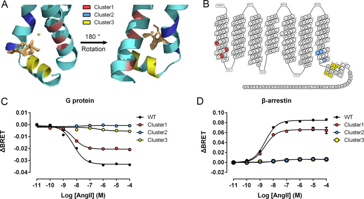
Fig. 9. Cluster mutations confirm P6 as an allosteric site.
A The positions of clusters 1 (red), 2 (blue), and 3 (yellow) on the intermediate structure (cyan). B The sites of alanine-scanning clusters on the 7TMs of the AT1 receptor. AngII-induced Gq activation (C) and β-arrestin 2 recruitment (D) through WT AT1 receptor (black) and cluster 1 (red), 2 (blue), and 3 (yellow) mutants. Three independent experiments were performed, and representative dose–response curves were shown. The bars indicate the mean ± SEM values.

