Figure 1.
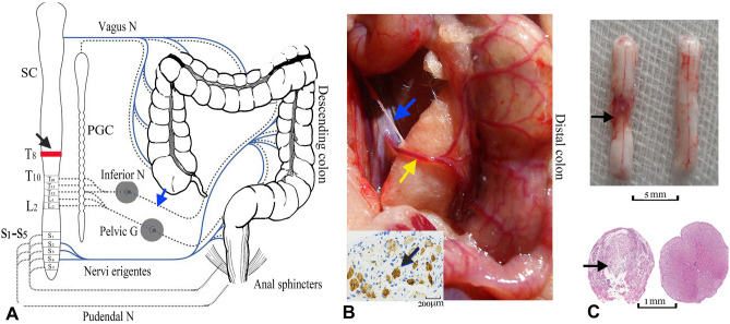
Surgery of SCI model and PSIMA. (A) Design of PSIMA for humans (the anatomy of colon), the sympathetic innervation (T10-L2) and parasympathetic innervation (S2–S4) are showed by dashed lines and blue lines, respectively. The blue arrow indicates the site of the nerve cut, and the black arrow indicates the level of the SCI. (B) The anatomy of accompanying sympathetic plexus and inferior mesenteric artery in a rat. The inferior mesenteric artery (indicated by yellow arrow), which originates from the anterior wall of the bifurcation of the left and right common iliac arteries, is mainly divided into the upper, middle, and lower branches, and with its accompanying sympathetic plexus (indicated by blue arrow) is situated to the distal colon. Lower left panel, an excised sympathetic nerve specimen stained with TH staining. (C) The upper panel showed the damaged segment of the spinal cord, four weeks after SCI (left) with the normal control (right), the lower panel, HE staining under the optical microscope, showed a complete damage at a cross section of the damaged segment. SC (spinal cord), PGC (paravertebral ganglia chain), Inferior N (inferior mesenteric ganglia), Pelvic G (pelvic ganglion), Vagus G (vagus ganglion), Pudendal G (pudendal ganglion), T8 and T10 (the 8th and 10th thoracic levels of the spinal cord), L2 (the second lumbar level of the spinal cord).

