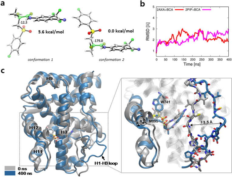
Figure 9.
Comparison of conformational changes of the wild type AR bound to BCA. (a) Two conformers of BCA with the lowest energy; Left: conformation 1 with the second lowest energy; Right: conformation 2 with the lowest in energy conformation. The dihedral angles of the four highlighted atoms (O-C-C-O) are shown. Images were created by using CYLView v.1.0.56156. (b) The backbone RMSDs of aMD simulations relative to the initial conformation. Gnuplot v5.257. (c) Comparison of the wild type ARs bound to BCA in conformation 2 at the initial (silver) and simulated structure after 400 ns (blue).Images were created by using VMD v1.9.352.

