Figure 3.
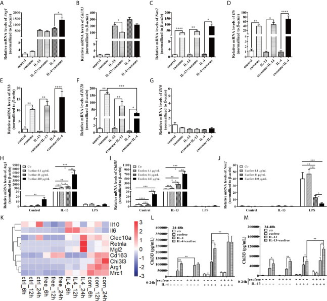
AC L4 exofree could promote a M2 polarization, while AC L4 exosomes induce M1 polarization of macrophage. (A–G) Macrophage activation markers Arg1 (A), Chi3l3 (B), Nos2 (C), and related interleukins Il6 (D), Il1b (E), Il12b (F), and Il10 (G) expression level were measured in the presence of AC L4 exosome, IL-4, IL-4+exosome, IL-13, and IL-13+exosome for 24 h using qPCR. The working concentration of AC L4 exosome is 25 μg/ml. The working concentration of IL-13 and IL-4 is 10 ng/ml. (H–J) Macrophage activation markers Arg1 (H), Chi3l3 (I), Nos2 (J) were measured in the presence of exofree, IL-4, IL-4+exofree, IL-13, and IL-13+exofree for 24 h using qPCR. (K) Signature gene expression profile of macrophage in the presence of PBS (ctr), free (AC L4 exofree), IL-4, com (IL-4+AC L4 exofree), and 6, 12, and 24 h represent 6, 12, and 24 h after the treatment, respectively. The working concentration of AC L4 exofree is 440 μg/ml. The working concentration of IL-13/IL-4 is 10 ng/ml. The working concentration of LPS is 0.05 μg/ml. The horizontal axis represents genes, and vertical coordinates represent types of treatment. (L, M) BMDMs were stimulated with PBS, AC L4 exofree, IL-4, IL-4+ AC L4 exofree, IL-13, IL-13+ AC L4 exofree in 0~24 h The culture medium was discarded. BMDMs were then washed with PBS and restimulated in 24~48 h as shown. And Chi3l3 protein level of 24~48 h culture medium was measured using ELISA. The working concentration of AC L4 exofree is 440 μg/ml. The working concentration of IL-13/IL-4 is 10 ng/ml. The detailed experiment information refer to Supplementary Table 2 . Data information: *P<0.05, **P<0.01, ***P<0.001, ****P<0.0001.

