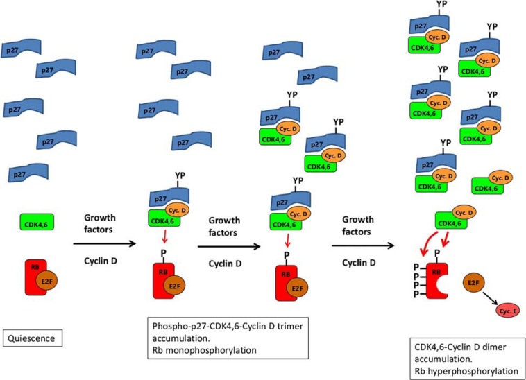
FIGURE 3.
A schematic illustrating a possible explanation of both RB-monophosphorylation and RB-hyperphosphorylation by CDK4,6/cyclin D. Growth factor stimulation of quiescent cells leads the expression of Cyclin D, which complexes with CDK4,6 and p27. The trimer, when phosphorylated on Tyr74 of p27 (marked YP) is a weak kinase for RB (resistant to palbociclib), enabling monophosphorylation of RB. (Whether tyrosine phosphorylation of p27 is restricted to the trimer or applies equally to the monomer, is not known. The lack of p27 monomers marked with YP in the diagram should not be taken therefore to imply that phosphorylation of the monomer does not occur.) As cyclin D expression continues, more and more of the trimer accumulates, eventually sequestering all the p27 monomers. From then on, further production of cyclin D leads to fully active CDK4,6/cyclin D dimers (sensitive to palbociclib) that hyperphosphorylate Rb. The derepressed E2F then induces expression of cyclin E which, together with CDK2, sustains Rb hyperphosphorylation thereafter.

