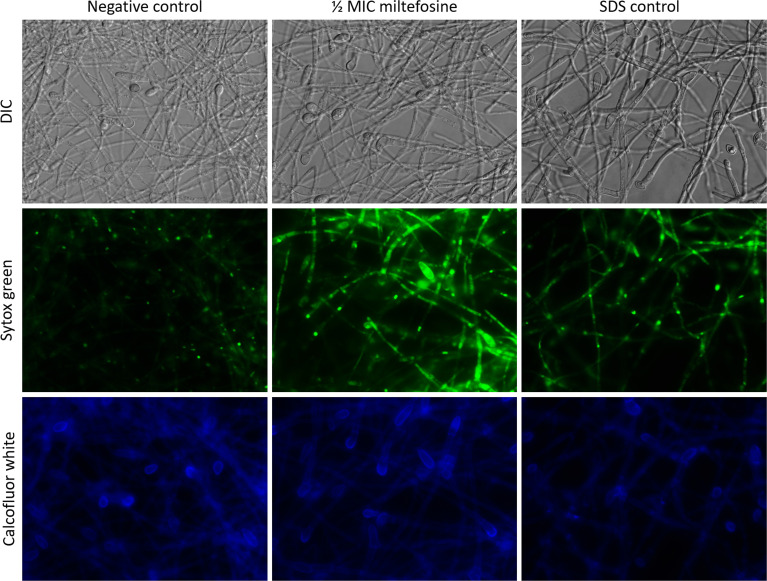
Figure 10.
Fluorescence microscopy of Scedosporium aurantiacum cells grown for 24 h at 37°C in the absence (negative control) or the presence of 0.5× MIC (2.0 μg/ml) of miltefosine. Sodium dodecyl sulphate (SDS) was used as a positive control of permeable membrane. Cells were stained with Sytox Green, which interacts with nucleic acid of permeable cells, and calcofluor white that stains the fungal cell wall. MIC, minimum inhibitory concentration.

