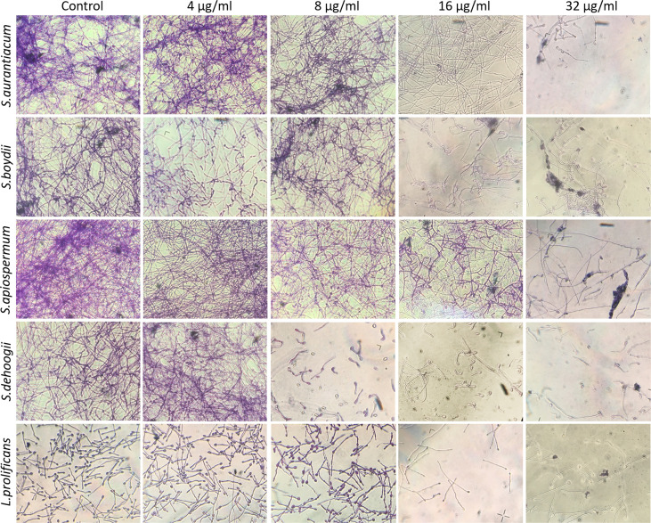
Figure 4.
Preformed biomass of Scedosporium aurantiacum, Scedosporium boydii, Scedosporium apiospermum, Scedosporium dehoogii and Lomentospora prolificans observed using a light microscope. Cells were grown in RPMI 1640 at 37°C for 24 h to form a fungal biomass and then a new 24-h incubation was performed in the absence (control) or the presence of 4, 8, 16 or 32 μg/ml of miltefosine. The fungal biomass was then stained with crystal violet and observed using a light microscope.

