FIGURE 4.
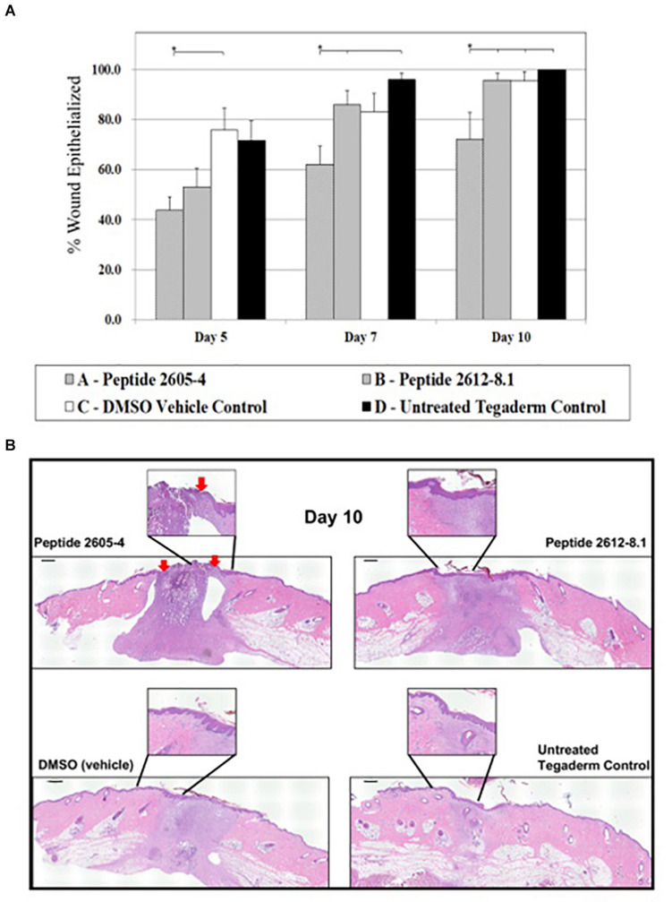
CLP 2605-4 treatment inhibited wound closure. (A) Graph showing percentage of epithelialization for all treatment groups at days 5, 7, and 10 post-treatment. The amount of re-epithelialized tissue was calculated as a percentage of the wounded area that was covered by newly formed epidermis. Treatment with CLP 2605-4 resulted in inhibition of epithelialization compared to other treatment groups (*p ≤ 0.05). (B) Representative wounds at day 10 stained with H&E are shown. Red arrows indicate extent of wound edges in CLP 2605-4 treated wounds indicating wound gap. Enlargements show epidermis fully covering wounds treated with CLP 2612-8.1, vehicle and untreated control wounds. Scale bar = 1000 μm.

