Figure 5.

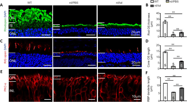
Luteolin (lut) preserves the morphological structure of photoreceptors and bipolar cells in rd10 mice.
(A, C, E) Images of rhodopsin (Green: Alexa Fluor-488, A), red/green opsin (red: Alexa Fluor-594, C), and PKC-α (red: Alexa Fluor-594, E) which stained the rod outer segment (OS), cone OS and rod bipolar cells, respectively, with DAPI (blue) staining in retinal sections at P26. Dotted lines in C and E illustrate the measured length of several cone OS and RBP dendrites, respectively. Scale bars: 20 μm in A and C, 10 μm in E. (B, D, F) Thickness of rod OS (B), length of cone OS (D), and RBP dendrites (F) in each group. Rod OS, cone OS, and bipolar dendrites were much thinner and shorter in the retinas of rd10 mice compared with WT controls, and luteolin increased these parameters at P26. Data are expressed as mean ± SEM. **P < 0.01 (one-way analysis of variance followed by Tukey’s post hoc test). Numbers within/near bars indicate quantities of mice tested. DAPI: 4′,6-Diamidino-2-phenylindole; INL: inner nuclear layer; IS: inner segment; ONL: outer nuclear layer; OPL: outer plexiform layer; OS: outer segment; P: postnatal day; PBS: phosphate-buffered saline; PKC-α: protein kinase C-α; RBP: rod bipolar cell; rd: rd10; WT: wild-type.
