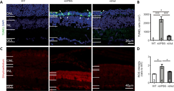
Figure 6.

Luteolin (lut) inhibites apoptosis and oxidative stress in the retinas of rd10 mice.
(A) Images of TUNEL (green) and DAPI (blue) staining of retinal slices at P26 (white arrowheads indicate TUNEL-positive cells). (B) Density of TUNEL-positive cells. In the WT retina, TUNEL-positive (apoptotic) cells were undetected. Apoptotic cells were increased in the retinas of rd10 mice, but were greatly decreased by luteolin. (C) Images of dihydroethidium (DHE, an indicator of ROS production) staining in retinal sections. Scale bars: 20 μm in A, 40 μm in C. (D) Fluorescent intensity of DHE staining across the whole retinal section normalized to the mean of WT controls. ROS levels were elevated in rd10 mice and further reduced by luteolin. Data are expressed as mean ± SEM. *P < 0.05, ***P < 0.001 (one-way analysis of variance followed by Tukey’s post-hoc test). Numbers within/near bars indicate quantities of mice tested. DAPI: 4′,6-Diamidino-2-phenylindole; GCL: ganglion cell layer; INL: inner nuclear layer; ONL: outer nuclear layer; P: postnatal day; PBS: phosphate-buffered saline; rd: rd10; ROS: reactive oxygen species; TUNEL: terminal deoxynucleotidyl transferase dUTP nick end labeling; WT: wild-type.
